
Dilute peptides lose more to vial and syringe surfaces
Peptides bind to glass and plastic, so dilute solutions lose a larger share of the dose. The data come from insulin bags and colistin labware, not home vials.

By Jay Spall, chemist and biochemist
Disclosure: Jay is a co-founder of The Peptide App. This review discusses the studies cited below; it is not a comprehensive live trial registry or treatment recommendation. Development and regulatory status can change. The app’s tools organize records and arithmetic and do not validate a research product.
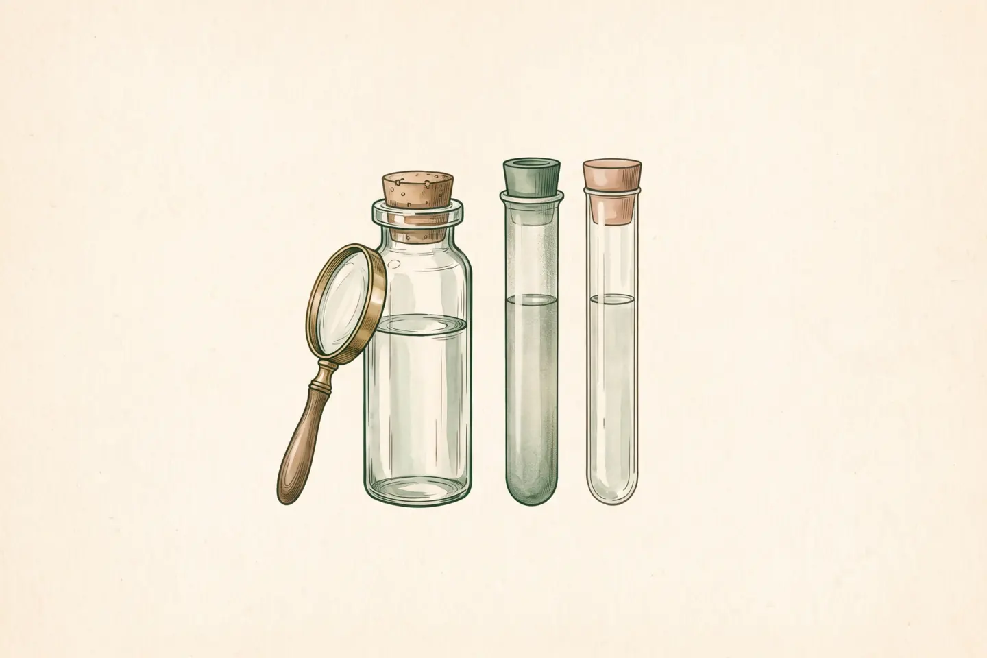
On this page
- Why do dilute peptides lose a bigger share to surfaces?
- How do drug makers keep dilute proteins off container walls?
- Does the container material change how much peptide is lost?
- Does diluting a peptide lower the dose recovered?
- Has peptide loss been measured in a home-reconstituted vial?
- How do you reduce peptide loss to vials and syringes?
- What is still unknown about peptide loss to surfaces?
- Sources
Key facts
| Question | Direct answer |
|---|---|
| Do peptides stick to vials and syringes? | Yes. Nonspecific binding of peptides and other small proteins to glass and plastic is documented for insulin in infusion bags and tubing [1] and for the lipopeptide colistin in glass and plastic labware [4]. |
| Does the container material matter? | Yes. Glass, polypropylene and polystyrene test tubes bound different amounts of colistin, and low-protein-binding plastic held onto less drug over time than standard tubes [4]. |
| Does a more dilute peptide solution lose a bigger percentage? | Yes. Surfaces have a roughly fixed binding capacity, so a more dilute insulin infusion bag gave lower recovery than a more concentrated bag of the same drug [1]. |
| Does it matter how long a dose sits in the syringe? | Yes. Surface loss builds over time instead of happening at once; colistin concentrations in some container materials fell substantially over 24 hours [4]. |
| Does the syringe marking equal the dose injected? | No. The barrel marking measures volume. Peptide the surface holds between draw and injection is invisible to that scale [2]. |
| How much peptide does a home-reconstituted vial lose? | Not established. None of the studies cited below drew a home-reconstituted research peptide into an insulin syringe and quantified how much reached the needle tip. |
5 sources cited. View sources
Why do dilute peptides lose a bigger share to surfaces?
A container wall binds a roughly fixed amount of peptide, so a dilute solution loses a larger fraction of its total dose. Glass and plastic are not inert to peptides in solution. Glass carries silanol groups that interact with charged and polar residues on a peptide backbone. Polypropylene and polystyrene present hydrophobic surfaces onto which peptides partition nonspecifically.
Tensiometry work on protein adsorption at low bulk concentrations shows that the amount lost to an interface depends heavily on the ratio of surface area to solution volume, and that the loss grows proportionally larger as bulk concentration drops [2]. That one relationship, surface area to volume, explains why dilution costs dose.
The binding sites inside a vial or syringe barrel work like a limited set of parking spots. A concentrated solution has so much peptide relative to the spots that only a small fraction parks. A dilute solution has far less peptide chasing the same spots, so a larger share of the dose ends up stuck to the wall.
How do drug makers keep dilute proteins off container walls?
Drug formulators add a carrier protein for the surface to bind instead. Formulation scientists add human serum albumin to botulinum toxin type A products to compete for nonspecific binding sites and keep the active protein in solution instead of on the container wall [3].
Some proteomics workflows apply the same carrier-protein logic to analysis, adding excess protein to small-volume samples to blunt surface losses during processing [5].
Does the container material change how much peptide is lost?
Container material changes the loss: colistin recovery differed across glass, polypropylene and polystyrene test tubes, and low-protein-binding microtubes lost less drug over 24 hours than standard tubes [4]. Colistin is a cyclic lipopeptide antibiotic, structurally closer to the small peptides people reconstitute at home than insulin is.
Researchers measured colistin directly in glass, polypropylene and polystyrene test tubes across concentrations of 0.125 to 8 mg/liter [4]. Starting recovery ranged from 44 to 102 percent of the expected concentration, depending on material. By 24 hours, recovery in some materials had fallen as low as 8 percent, with loss half-lives of 0.9 to 12 hours across materials [4].
Low-protein-binding microtubes performed better. They held 59 to 90 percent of the starting concentration at 24 hours, while other materials dropped to single digits [4].
These are in vitro labware data, not a home-dosing trial. They are still a direct, quantitative demonstration of surface loss, in a molecule class close enough to home-use peptides to matter.
Does diluting a peptide lower the dose recovered?
Dilution lowers recovery: a 3 unit per 250 mL insulin infusion bag returned a smaller share than a 45 unit per 250 mL bag of the same insulin [1]. Recovery from the bag itself was 29.7 percent at the lower concentration and 37.3 percent at the higher one. After the fluid ran through standard administration tubing, recovery fell to 11.9 percent and 30.6 percent [1].
The comparison used the same insulin, the same bag type and the same tubing. Starting concentration alone drove a roughly threefold difference in final recovery.
Insulin is not the peptide in a research vial, and IV bags and tubing do not share the geometry of a reconstitution vial and a syringe barrel. The insulin comparison is still the cleanest available demonstration that diluting the same drug in the same container class predictably worsens recovery. Standard peptide guides never make that point.
Syringe markings do not capture the loss either. The number on the barrel is a volume marker, and peptide the surface holds between draw and injection is invisible to that scale [2]. The difference between syringe units and dose explains what those markings do measure.
Has peptide loss been measured in a home-reconstituted vial?
None of the studies cited below followed a research peptide reconstituted at home into a specific insulin syringe and quantified how much reached the needle tip. Surface adsorption itself is well established and cross-validated across at least two distinct drug classes and multiple container materials [1][2][4].
The colistin and insulin results are strong, cross-molecule laboratory evidence that the mechanism operates and scales in the directions described. That surface loss applies, at some magnitude, to a peptide reconstituted at home is highly plausible on mechanistic grounds. It is consistent with the carrier-protein strategies pharmaceutical formulators build for this exact problem [3][5].
What is not established is the specific percentage lost from any one person's vial and syringe. Anyone quoting a precise number for a home preparation is extrapolating past what these data support.
How do you reduce peptide loss to vials and syringes?
Keeping the reconstituted concentration reasonably high and injecting soon after drawing both follow directly from the surface-loss data.
- Keep the concentration reasonably high. More diluent makes a more dilute peptide, which loses proportionally more to the vial wall. The same logic separated 29.7 percent from 37.3 percent recovery in the insulin bag comparison [1]. A reconstitution ratio chosen only to make the syringe math convenient works against the dose; choosing a reconstitution volume for syringe accuracy covers the other side of that trade-off.
- Inject promptly after drawing. Time in the syringe is not free. The colistin data show loss accumulating over hours, not at the moment of contact [4]. Drawing up a dose and injecting promptly is more defensible than drawing it up and letting it sit.
- Treat low-binding syringes as unproven. Low-protein-binding plastics measurably outperformed standard polystyrene and polypropylene tubes in the colistin work [4]. None of these studies measured whether that advantage carries over to a specific syringe brand's barrel coating. "Buy a low-binding syringe" is a plausible extension of documented material differences, not a proven fix for any particular equipment.
- Judge a cloudy vial separately. Surface adsorption data do not address visual assessment of contamination or precipitation. A cloudy vial needs its own reasoning, not a blanket rule; what cloudiness in an injectable vial means covers that question.
What is still unknown about peptide loss to surfaces?
The exact amount of peptide missing from a home-prepared dose is unknown. The cited studies leave three related questions open:
- Desorption. Whether adsorbed peptide desorbs back into solution over time is unanswered.
- Saturation. Whether repeated draws from the same vial progressively saturate the available binding sites, and so reduce further loss, is unknown.
- Home-use peptides. How surface loss plays out for the peptide classes reconstituted at home, rather than insulin or colistin, has not been worked out.
The mechanism is real, and the direction of every effect described is consistent. Neither the published adsorption studies nor a certificate of analysis can give the exact number missing from a dose.
Sources
Last updated

Written by
Chemist and biochemist. Co-founder and author, The Peptide App.
Jay is a chemist, biochemist and entrepreneur whose work connects scientific research with consumer health products. He has held Chief Science Officer and product development leadership roles and previously served as Chief Revenue Officer at Minicircle.
Keep reading
Storage and handling
Vial cloudiness has at least four causes; two are quality failures
Vial cloudiness can mean air, undissolved solid, aggregation, or contamination, and only some warrant discarding. Clarity proves neither sterility nor potency.
Storage and handling
Peptide gels in the vial come from self-assembly, not chemical damage
Peptide gelling in the vial is a physical rearrangement of intact chains, not chemical damage. No PubMed study shows that AOD-9604 or kisspeptin-10 gels.
Reconstitution and syringe math
Peptide reconstitution volume should put your dose mid-syringe
Reconstitution volume changes peptide concentration, not the amount in the vial. Pick a volume that puts your dose mid-barrel to reduce measurement error.
Profiles and tools

The Peptide App
Track protocols, doses, and reconstitution in one place.
Save your calculations, set reminders, log doses, and keep outcome notes — free to start.
 or scan the QR code →